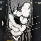
Figure 2.
Anterior Laparoscopic Approach Combined with Posterior Approach for Lumbosacral Neurolysis: A Case Report
Sheng Wang#, Nan Lu, Yongchuan Li, Xiaohuang Tu* and Aimin Chen*
Published: 25 November, 2024 | Volume 8 - Issue 1 | Pages: 010-013

Figure 2.:
The patient's preoperative evaluation and intraoperative neurolysis. (A-C) The area of sensory disturbance before operation, the area with the red border is the senseless area, and the area with the black border is the sensory numbness area. (D-E) X-ray and CT three-dimensional reconstruction before operation. (F) Posterior approach lumbosacral neurolysis, the yellow border area is the loosened nerve. (G) Laparoscopic exposure of the lumbar nerve, the yellow border area shows the iliac vessels and the purple border area is the lumbar nerve. (H) Laparoscopic exposure of the lumbar nerve, the yellow border area is the loosened nerve. (I and J) Six months after surgery, the abdominal and lumbar incisions recovered well. (K) Six months after surgery, the motor function of the patients was obviously improved.
Read Full Article HTML DOI: 10.29328/journal.aceo.1001020 Cite this Article Read Full Article PDF
More Images
Similar Articles
-
Anterior Laparoscopic Approach Combined with Posterior Approach for Lumbosacral Neurolysis: A Case ReportSheng Wang#,Nan Lu,Yongchuan Li,Xiaohuang Tu*,Aimin Chen*. Anterior Laparoscopic Approach Combined with Posterior Approach for Lumbosacral Neurolysis: A Case Report. . 2024 doi: 10.29328/journal.aceo.1001020; 8: 010-013
Recently Viewed
-
Characteristics of Stones Ageing for Climate Resilience Due to Carbon Lifeform EnvironmentSolomon I Ubani*. Characteristics of Stones Ageing for Climate Resilience Due to Carbon Lifeform Environment. Ann Civil Environ Eng. 2024: doi: 10.29328/journal.acee.1001069; 8: 063-069
-
Air Quality Dynamics in Sichuan Province: Sentinel-5P Data Insights (2019-2023)Hossam Aldeen Anwer and Abubakr Hassan*. Air Quality Dynamics in Sichuan Province: Sentinel-5P Data Insights (2019-2023). Ann Civil Environ Eng. 2024: doi: 10.29328/journal.acee.1001068; 8: 057-062
-
The Influence of Gravity on the Frequency of Processes in Various Geospheres of the Earth. Biogenic and Abiogenic Pathways of Formation of HC AccumulationsAA Ivlev*. The Influence of Gravity on the Frequency of Processes in Various Geospheres of the Earth. Biogenic and Abiogenic Pathways of Formation of HC Accumulations. Ann Civil Environ Eng. 2024: doi: 10.29328/journal.acee.1001067; 8: 052-056
-
Review of AI in Civil EngineeringVinayak Hulwane*. Review of AI in Civil Engineering. Ann Civil Environ Eng. 2024: doi: 10.29328/journal.acee.1001066; 8: 048-051
-
The Effect of Cellulose Fiber on the Bending Strength of Autoclaved Aerated ConcreteSvitlana Lapovska, Nicholas Chernenko, Mykola Konoplya. The Effect of Cellulose Fiber on the Bending Strength of Autoclaved Aerated Concrete. Ann Civil Environ Eng. 2024: doi: 10.29328/journal.acee.1001065; 8: 045-047
Most Viewed
-
Evaluation of Biostimulants Based on Recovered Protein Hydrolysates from Animal By-products as Plant Growth EnhancersH Pérez-Aguilar*, M Lacruz-Asaro, F Arán-Ais. Evaluation of Biostimulants Based on Recovered Protein Hydrolysates from Animal By-products as Plant Growth Enhancers. J Plant Sci Phytopathol. 2023 doi: 10.29328/journal.jpsp.1001104; 7: 042-047
-
Sinonasal Myxoma Extending into the Orbit in a 4-Year Old: A Case PresentationJulian A Purrinos*, Ramzi Younis. Sinonasal Myxoma Extending into the Orbit in a 4-Year Old: A Case Presentation. Arch Case Rep. 2024 doi: 10.29328/journal.acr.1001099; 8: 075-077
-
Feasibility study of magnetic sensing for detecting single-neuron action potentialsDenis Tonini,Kai Wu,Renata Saha,Jian-Ping Wang*. Feasibility study of magnetic sensing for detecting single-neuron action potentials. Ann Biomed Sci Eng. 2022 doi: 10.29328/journal.abse.1001018; 6: 019-029
-
Pediatric Dysgerminoma: Unveiling a Rare Ovarian TumorFaten Limaiem*, Khalil Saffar, Ahmed Halouani. Pediatric Dysgerminoma: Unveiling a Rare Ovarian Tumor. Arch Case Rep. 2024 doi: 10.29328/journal.acr.1001087; 8: 010-013
-
Physical activity can change the physiological and psychological circumstances during COVID-19 pandemic: A narrative reviewKhashayar Maroufi*. Physical activity can change the physiological and psychological circumstances during COVID-19 pandemic: A narrative review. J Sports Med Ther. 2021 doi: 10.29328/journal.jsmt.1001051; 6: 001-007

HSPI: We're glad you're here. Please click "create a new Query" if you are a new visitor to our website and need further information from us.
If you are already a member of our network and need to keep track of any developments regarding a question you have already submitted, click "take me to my Query."


